②SATのUS画像は、びまん性に不均一な実質像と局所性の著明な低エコー領域(結節様)であり、カラードプラでは血流の低下が基本的な特徴所見である。
SATのBモードエコー画像は、びまん性の不均一な実質像と著明な低エコー域の存在を特徴とする(2)。特に楕円形の境界不明瞭な低エコー域の存在は典型的と言われている(14,15)。様々な形をした不明瞭な不均一域は”lava-flow“(溶岩流)エコーパターンと呼ばれている。時には結節状で癌との鑑別が困難な所見を示す場合がありFNABを施行される場合もある(16)。
SATのエコー所見は様々であるが、human leukocyte antigen(HLA)と関連し、HLA-B*18:01の存在はエコー所見に変化を与える決定因子と考えられ、典型的な画像所見はHLA-B*18:01が存在するかどうかで決定される傾向があると言われている(4)。HLA-B*35の共存は典型所見に修飾を加える形で作用する可能性を疑われている。
一方color doppler所見は、著明に血流低下を示し著明な血流亢進を示すGraves病との鑑別に利用されている(17)。
[Case Presentaion]
[CC]全身倦怠感と動悸、夜間の発熱(38℃)
[PH]6月の初旬、咽頭痛、輪状軟骨付近の圧痛と39度台の熱発で耳鼻科受診、ペニシリン製剤等の投薬治療を受けたが改善せず。
[PE] BT 36.4℃, BP 123/77, P 104bpm, SpO2=97% 甲状腺左葉に圧痛あり
[Blood Chemistry]
WBC 10’600/μL(Neu 83%), CRP 6.34 mg/dL, ASO 197 IU/mL
T.Bil 0.6 mg/dL, AST 86 U/L, ALT 128 U/L, GGT 149 U/L, ALP 139 U/L, CHE 329 U/L, BUN 10.8 mg/dL, CR 0.65 mg/dL, Na 140 mEq/L, K 4.3 mEq/L, Cl 102 mEq/L, TSH(ECLIA) LT0.005 μIU/mL, FT3 11.35 pg/mL(2.30〜4.00), FT4 4.88 ng/dL(0.90〜1.70), TRAb 結合阻害率42.0%(-10.0〜10.0%)
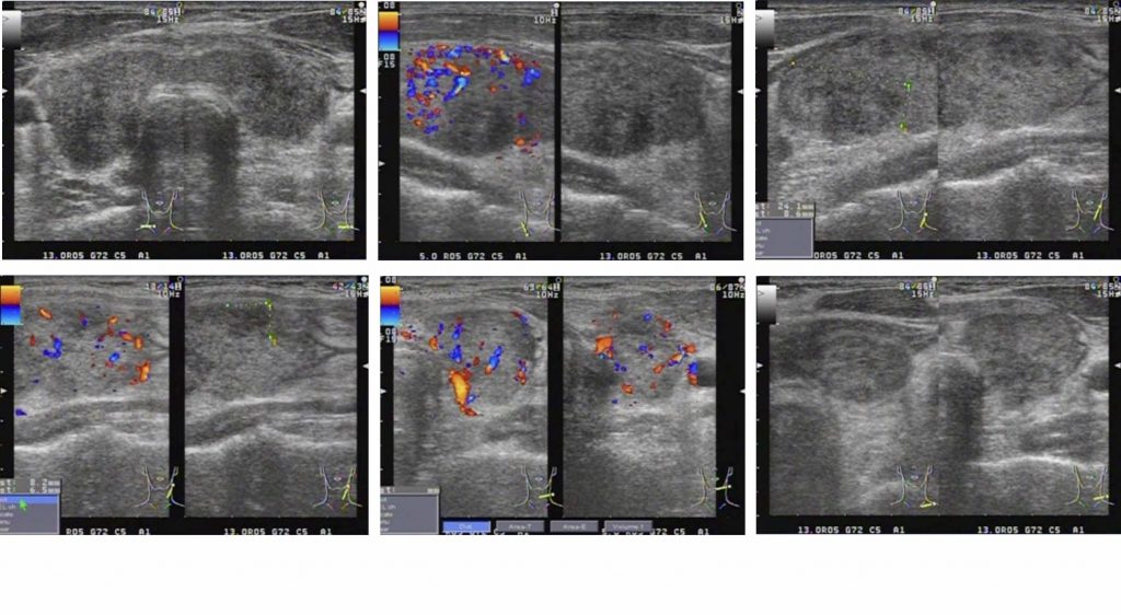
[US & Color doppler findings] びまん性に不均一で粗い実質エコーを呈し表面はやや凸凹調である。甲状腺内に境界不明瞭な低エコー領域(一部結節様)を認める。SATを疑う所見であるが、カラードプラ所見は通常のSATとは異なり、実質の血流増加が認められる。

[Clinical Course]
SATと考暫定診断し、アセトアミノフェンを4日程投与したが発熱、頸部痛の改善が乏しいためNSAID(ナイキサン)を処方したところ症状は改善した。しかしながらTRab陽性のためSATの再燃などの可能性が懸念され基幹病院専門医に精査を依頼した。発熱、頸部痛消失後もFT3高値が持続、2年間follow upいただいたところでTRAb陰性化、自然軽快された。SAT→Graves発症ではなく、GravesにSATが併発した症例と診断された。
[Final Diagnosis]
SAT on Graves disease : バセドウ病に併発した亜急性甲状腺炎
Uploaded on April 02, 2021.
参考文献
- De Quervain F. Mitteilungen aus den Grenzgebieten der Med- izin und Chirurgie. In: Die akute, nicht eiterige Thyreoiditis und die Beteiligung der Schilddrüse an akuten Intoxikationen und Infektio- nen überhaupt. Jena Germany: G. Fischer; 1904:1-165.
- Li JH, Daniels GH, et al. Painful subacute thyroiditis commonly misdiagnosed as suspicious thyroid nodular disease. Mayo Clin Proc Inn Qual Out 2021: 1-8
- Ohsako N, Tamai H, Sudo T, et al. Clinical characteristics of subacute thyroiditis classified according to human leukocyte an- tigen typing. J Clin Endocrinol Metab 1995;80(12):3653-3656.
- Stasiak M, Tymoniuk B, et al. Sonographic pattern of subacute thyroiditis is HLA-dependent. Front Endocrinol 2019; 10(3): 1-8
- Brancatella A, Ricci D, et al. Subacute thyroiditis after Sars-COV2 infection. J Clin Endocrinol Metab 2020; 105(7): 1–4
- Nishihara E, Ohye H, et al. Clinical characteristics of 852 patients with subacute thyroiditis before treatment. Inter Med 2008; 47: 725-729.
- Volpé RC. Subacute (non-suppurative) thyroiditis. In: Werner SC (ed) The Thyroid 4th edition. Harper & Row Publishers, Inc. Maryland 1978: 986-988.
- Sriphrapradang C, Bhasipol A. Differentiating Graves’ disease from subacute thyroiditis of serum free triiodothyronine to free thyroxine. Ann Med Surg 2016; 10: 69-72
- Sümbül HE, Acıbucu F. Graves’ disease and thyroiditis can be differentiated using only free thyroid hormone levels. Eur Res J 2019; 0(0): 1-5
- Iitaka M, Momotani N, et al. TSH receptor anti- body-associated thyroid dysfunction following subacute thyroiditis. Clin Endocrinol 1998; 48: 445-453.
- Kageyama K, Kinoshita N, et al. A Case of Thyrotoxicosis due to Simultaneous Occurrence of Subacute Thyroiditis and Graves’ Disease. Case Reports in Endocrinol 2018; Article ID 3210317, 3 pages
- Fukushima A, Tanabe M et al. A rare case of subacute thyroiditis simultaneously complicated by Graves’ disease: A case report and review of the literature. J Endocrinol Thyroid Res 2017; 2(4): 1-5
- Nakano Y, Kurihara U, et al. Graves’ disease following subacute thyriditis. Tohoku J Exp Med 2011; 225: 301-309
- Park SY, Kim EK, et al. Ultrasonographic characteristics of subacute granulomatous thyroiditis. Korean J Radiol 2006; 7: pp229-pp234
- Sencar ME, Calapkulu M, et al. The contribution of ultrasonographic findings to the prognosis of subacute thyroiditis. Arch Endocrinol Metab 2020; 64(3): 306-331
- Lee MYW, Lam WWC, et al. Subacute thyroiditis-an often overlooked sonographic diagnosis. Report of 3 cases. J Ultrasound Med 2016; 35: 1095–1100
- Frates MC, Marqusee E, ea al. Subacute granulomatous (de Quervain) thyroiditis. Grayscale and color doppler sonographic characteristics. J Ultrasound Med 2013;32: 505-511